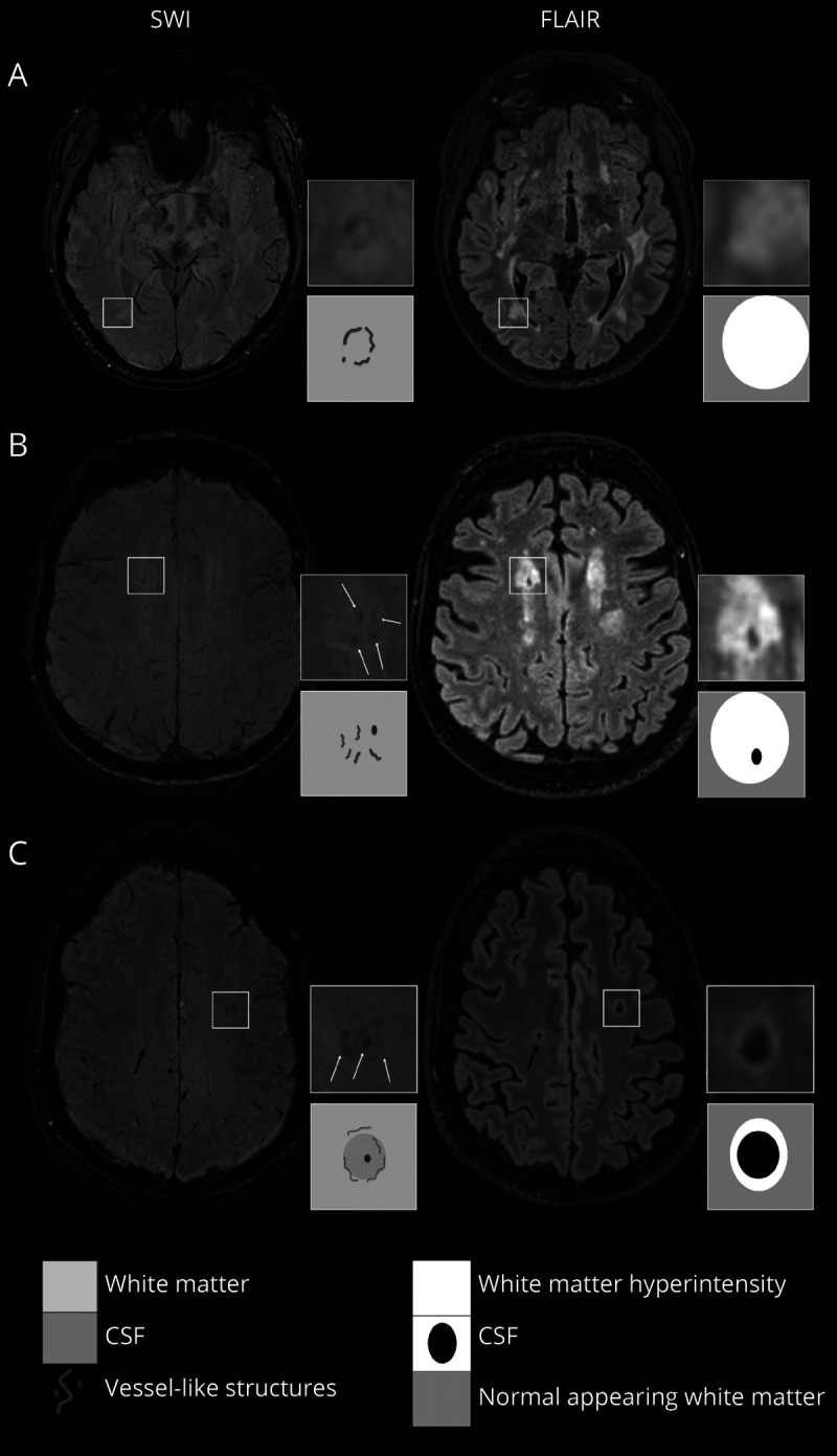
Figure 1. Vessel-Clusters on Susceptibility-Weighted Imaging and Corresponding Appearance on Fluid-Attenuated Inversion Recovery.

Vessel-clusters indicated in squares have been augmented (4x) to show details of susceptibility-weighted imaging (SWI) and fluid-attenuated inversion recovery (FLAIR) sequences, another not augmented vessel-cluster is indicated with a black arrow (C). For each patient (A with CADASIL, B and C with sporadic SVD), we present the most informative SWI (left) and corresponding FLAIR (right) sequences to show vessel-cluster appearance. Vessel-clusters are characterized by clustered small tubular structures, visible as low-signal dots (B and C) or lines (A, B, and C) on SWI not corresponding to normal appearing deep medullary veins (visible as mild low-signal parallel lines reaching the lateral ventricles from the deep white matter). Vessel-clusters are seen within white matter hyperintensities with different grades of cavitation: none (A), partial (B), or complete (C) cavitation. The schematic appearance of vessel-clusters on SWI and grade of cavitation on FLAIR are represented in the corresponding squares below the augmentations. SVD = small vessel disease.
