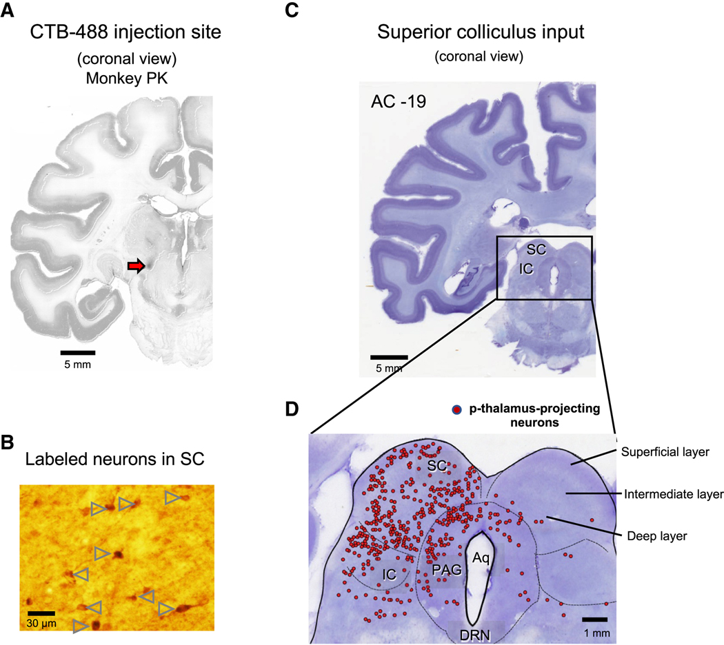
Figure 2. Superior colliculus projects to the posterior thalamus.
A. Injection site of retrograde tracer. We injected cholera toxin subunit B conjugated with Alexa Fluor-488 (CTB-488) into the region of the p-thalamus where the marking lesion was made (Red arrow). Slices were stained by anti-Alexa Fluor 488 antibody to detect the CTB-488 with high sensitivity. B. Retrogradely labeled neurons in the superior colliculus (SC). Arrow heads indicate the SC neurons projecting to the p-thalamus labeled by anti-Alexa Fluor 488 antibody. C. Coronal view of brain stem including the SC, inferior colliculus (IC), and periaqueductal grey (PAG). AC: Anterior commissure. D. Retrogradely labeled neurons in the SC, IC, and PAG. Red dots indicate the p-thalamus-projecting neurons in the SC, IC, and PAG.
See also Figure S1.

