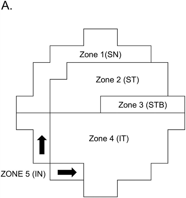
Figure 1.

Visual field zones of 10-2 VF devised by Hood et al.13 Note that this map assumed 5 distinct VF zones based on their vulnerability to damage in the macula. Zone 1 = superior nasal zone, Zone 2 = superior temporal zone, Zone 3 = superior temporal band, Zone 4 = inferior temporal zone, Zone 5 = inferior nasal zone.
