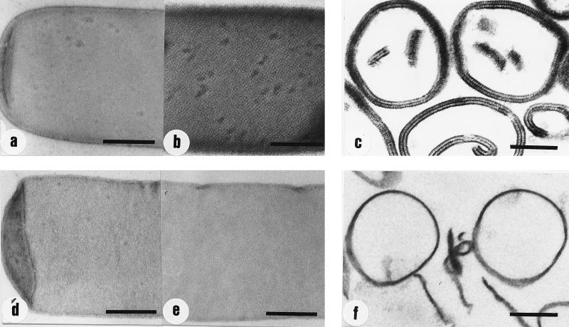
FIG. 2.
Electron micrographs of negatively stained and ultrathin-sectioned preparations of native and HF-extracted peptidoglycan-containing sacculi of B. sphaericus CCM 2177. (a and b) Negatively stained preparations of native peptidoglycan-containing sacculi before and after recrystallization of the S-layer protein, respectively; (c) ultrathin-sectioned preparation of native peptidoglycan-containing sacculi after recrystallization of the S-layer protein; (d and e) negatively stained preparations of HF-extracted peptidoglycan-containing sacculi (48% HF, 48 h at 4°C) before and after the addition of the GHCl-extracted S-layer protein and dialysis, respectively; (f) ultrathin-sectioned preparation of HF-extracted peptidoglycan-containing sacculi after the addition of the GHCl-extracted S-layer protein and dialysis. Bars, 250 nm.

