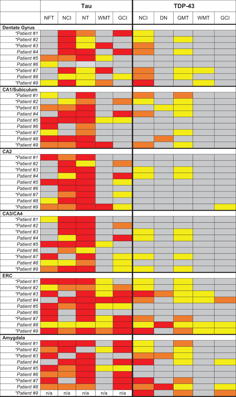
Table 2.
Neuropathological Evaluation of Tau (CP-13) and TDP-43 Proteinopathy Burden in Medial Temporal Lobe Regions


|
Each region was rated semi-quantitatively by a neuropathologist based on the type of cellular inclusion, morphology, and burden of pathology identified by immunohistochemistry (gray = Not Identified, yellow = mild/sparse, orange = moderate, red = severe/frequent). W scores reflecting volume loss based on antemortem T1-weighted magnetic resonance imaging are provided for the hippocampus, entorhinal cortex, amygdala, and a composite medial temporal lobe (MTL) region. Lower W scores represent lower gray matter volume in the region of interest compared with a large group of clinically normal, cognitively healthy controls.
NFT, neurofibrillary tangles; DN, dystrophic neuritis; GMT, gray matter threads/dots; WMT, white matter threads/dots; GCI, glial cytoplasmic inclusions; NCI, neuronal cytoplasmic inclusions (other than tangles or Pick bodies); NT – neuropil threads, ERC, entorhinal cortex.
Patients with chronic traumatic encephalopathy (CTE) pathology at autopsy (High CTE - #1, #2, #7, #9; Low CTE - #3).
