Figure 2.

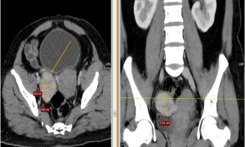
Contrast-enhanced CT shows a relatively well-defined isohypodense soft tissue mass lesion showing post-contrast enhancement measuring 5.2×4.4×4.2 cm (AP-TR-CC) in the right adnexa with normal left ovary, Fallopian tubes and uterus.

Contrast-enhanced CT shows a relatively well-defined isohypodense soft tissue mass lesion showing post-contrast enhancement measuring 5.2×4.4×4.2 cm (AP-TR-CC) in the right adnexa with normal left ovary, Fallopian tubes and uterus.