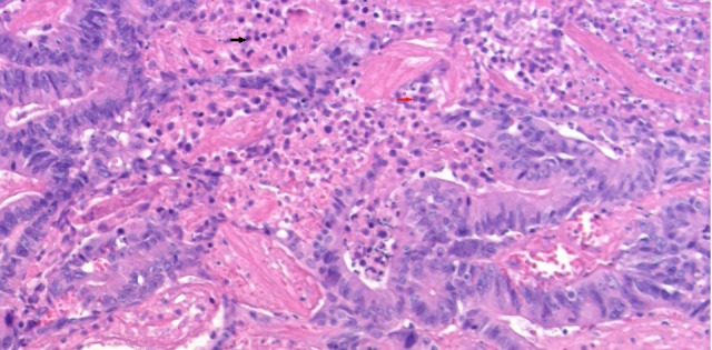
Figure 3.

Histopathology of excised right ovarian tissue shows polygonal cells having distinct cell borders, central nuclei with moderate eosinophilic cytoplasm, without necrosis/haemorrhage. Stroma is oedematous at places with loosely dispersed tumour cells with dilated thin-walled blood vessels—features suggestive of Sertoli-Leydig cell ovarian tumour (Sertoli cell indicated by red arrow, Leydig cell marked by black arrow).
