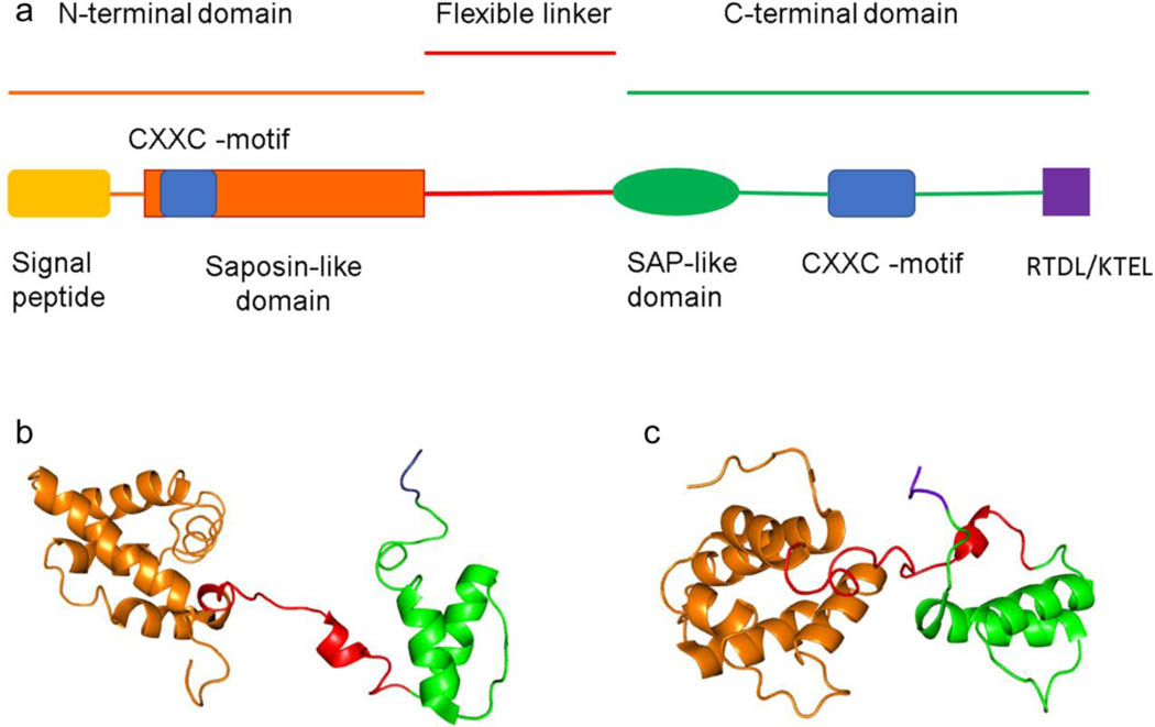
Figure 1. Structure of human (Homo sapiens) MANF and CDNF.
The different domains and motifs of MANF and CDNF structure. “RTDL” and “KTEL” are the ER retention sequences for MANF and CDNF, respectively (A). The structures of MANF (B) and CDNF (C) created from PDB codes 2KVD (residues 25–182) and 4BIT (residues 27–187), respectively, using PyMOL 2.3.4 software (Parkash et al., 2009; Latge et al., 2015).

