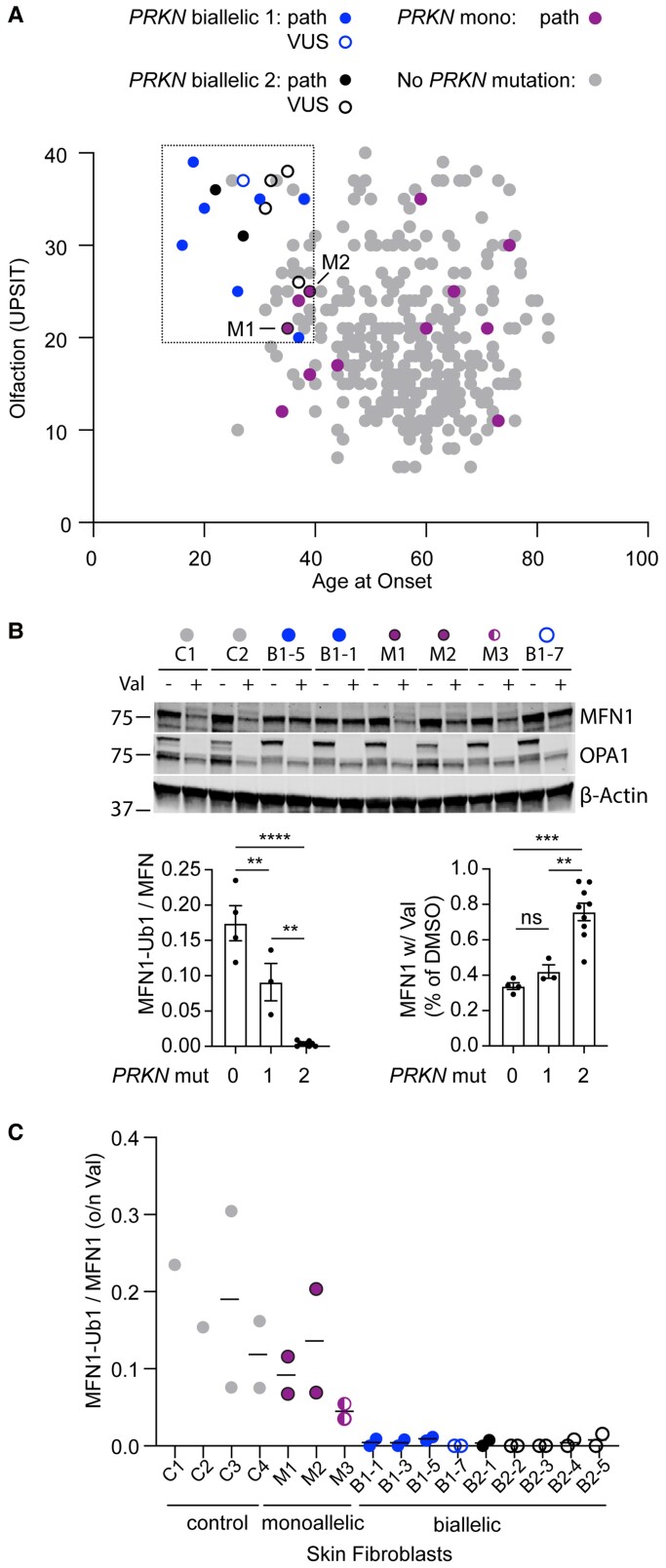
Figure 2.
Biallelic and monoallelic PRKN mutation carriers can be distinguished by their phenotype and functional assays in their fibroblasts. (A) Scatterplot depicts scores from smell identification test (UPSIT, y-axis) and age-at-onset (x-axis) for genotyped Parkinson disease patients in the NIH-PD cohort with no PRKN mutations (grey), one PRKN mutation (magenta), or two PRKN mutations (blue). A second group of patients with two PRKN mutations (black) was identified outside of the consecutive series. Those with a novel variant or VUS are shown as open symbols. Two patients with one PRKN mutation and whose fibroblasts were evaluated in C have a black border. (B and C) Patient fibroblasts were treated with vehicle (DMSO) or 10 mM of valinomycin (val) overnight (o/n), separated on SDS-PAGE gels, and immunoblotted for the PARKIN substrate MFN1, as shown in representative blot. The ratio of MFN-Ub1 (corresponding to the slower migrating band) to MFN1 was calculated for each cell line treated with val. Each cell line was assayed in two biological replicates with exception of lines C1 and C2, which were assayed in one replicate each. All cell lines from PRKN carriers were from individuals with Parkinson’s disease, except for M3, who was the unaffected sibling of B1-7. Graphs of the summary data are shown on the bottom. (C) Individual data for experiment described in B.

