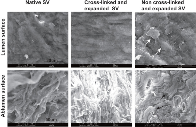
Fig. 3.
Scanning electron microscopy of human saphenous vein (SV). The ultrastructure of luminal and abluminal surfaces was observed in SVs with different processing (luminal surface magnification, ×4000; abluminal surface magnification, ×2400). The white arrows show patches of endothelium containing denuded and curly cells. The black arrows show fibre fracture.

