
Figure 5.
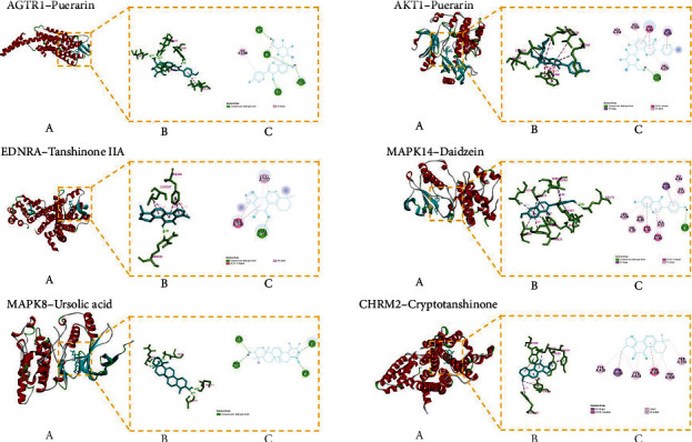
The molecular docking pattern. Each target protein with its active ingredient molecular docking pattern diagram is divided into three little figures (a), (b), and (c). (a) Represents the overall binding position of small molecule compounds (sticks model) on related proteins (cartoon model). (b) Shows the interaction of small molecule compounds (blue) with important residues (green) on related proteins. (c) Displays a two-dimensional plan, which is convenient for observing the hydrogen bonding and hydrophobic interactions between small molecule compounds and protein residues.