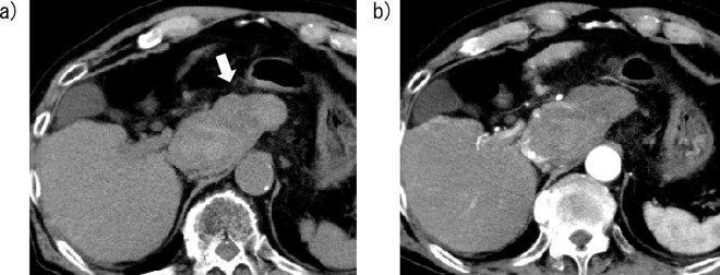
Figure 4.
Computed tomography (CT) images of Case 2 obtained five days after lenvatinib initiation. a) A nonenhanced CT image shows a high-attenuation area around the caudate lobe, indicating a hematoma caused by hepatocellular carcinoma rupture (arrow). b) A contrast-enhanced CT image shows a necrotic area in the HCC nodule in the caudate lobe.

