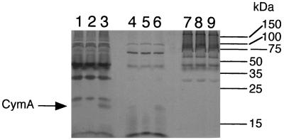
FIG. 2.
Heme-stained SDS-PAGE profiles of subcellular fractions prepared from S. putrefaciens cells grown anaerobically with TMAO as the electron acceptor. The lanes were loaded with 50 μg of protein of each of the following subcellular fractions: cytoplasmic membrane (lanes 1 to 3), soluble fraction (lanes 4 to 6), and outer membrane (lanes 7 to 9). Fractions were prepared from MR-1/pVK100 (lanes 1, 4, and 7), MR1-CYMA/pVK100 (lanes 2, 5, and 8), and MR1-CYMA/pCMTN1-VK (lanes 3, 6, and 9). The bars and numbers at the right indicate the migration and masses of the protein standards.

