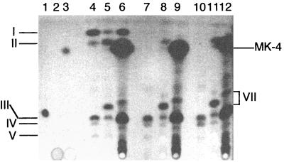
FIG. 7.
Thin-layer chromatogram of quinone standards and of quinones isolated from S. putrefaciens cells grown anaerobically with fumarate as the electron acceptor and supplemented with either DHNA or MK-4. Standards were loaded in lanes 1 to 3 as follows: lane 1, ubiquinone-10; lane 2, DHNA; lane 3, menaquinone-4. The other lanes were loaded with quinone extracts (equivalent to 0.18 g of wet cells) isolated from cells of the following strains: lanes 4 to 6, MR-1; lanes 7 to 9, MR-1A; lanes 10 to 12, MR-1B. During cell growth, the medium was supplemented with 10 μM DHNA (lanes 5, 8, and 11), 10 μM MK-4 (lanes 6, 9, and 12), or the solvent for these quinones as a control (lanes 4, 7, and 10). The samples were loaded at the bottom, and migration was upward. As described above for MR-1, spots I and II are methylmenaquinone and menaquinone, respectively, and spots III, IV, and V are ubiquinones. The prominent presence of MK-4 in cells grown in the presence of MK-4 is indicated at the right. The DHNA standard (15 nmol; lane 2) was not visually detectable under these conditions. The spectra for the spots indicated by VII (not shown) were consistent with ubiquinone.

