Fig. 1.
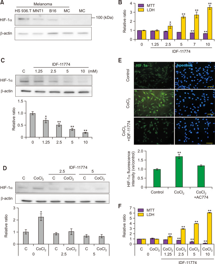
IDF-11774 reduces CoCl2-induced HIF-1α upregulation in cultured B16F10 melanoma cells. (A) Western blot analysis for HIF-1α protein expression in three kinds of cultured melanoma cells. (HS936T, MNT1, and B16F10) and primary cultured normal human epidermal melanocytes (MC). (B, C) MTT assay for cell viability and LDH release for cytotoxicity (B) and Western blot analysis for HIF-1α protein expression in B16F10 cells treated with different concentrations (0, 1. 25, 2.5, 5, and 10 mM) of IDF-11774 for 48 h. (D, E) Western blot analysis for relative ratios of HIF-1α levels (D) and representative immunofluorescent staining using anti-HIF-1α antibody in B16F10 treated with IDF-11774 in the absence or presence of CoCl2 for 48 h. Nuclei were counter-stained with Hoechst 33258 (Bar=0.05 mm) (E). (F) MTT assay and LDH release in B16F10 treated with IDF-11774 in the absence or presence of CoCl2 for 48 h. β-Actin was used as an internal control for Western blot analysis. Intensities of immunofluorescence staining were measured using a Wright Cell Imaging Facility ImageJ software. Data in the graph represent mean ± SD of relative values compared to non-treated control from four independent experiments. *p<0.05, **p<0.01.

