Fig. 5.

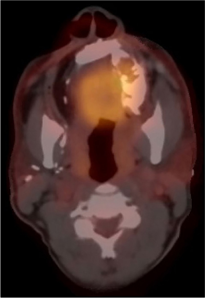
Two years follow up: PET/CT* imaging revealed evidence of disease in the left maxilla after two years from the excisional biopsy. (*Positron Emission Tomography—Computed Tomography)

Two years follow up: PET/CT* imaging revealed evidence of disease in the left maxilla after two years from the excisional biopsy. (*Positron Emission Tomography—Computed Tomography)