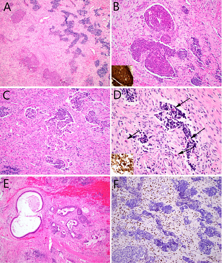
Fig. 1.
Case 1 consisted of nests of round cells, set in a fibrous stroma with a few dilated vessels (A). Some of the tumor nests were squamous and showed nuclear beta-catenin immunostaining (inset) (B), but most of them were made up of primitive cells with high nuclear:cytoplasmic ratios and foci of glio-fibrillary stroma (C). Scattered rhabdomyoblasts (arrows) that were desmin positive (inset) were noted within the round cell and spindle cell tumor components (D). There were numerous benign-appearing invaginations of surface epithelium, many of which were cystic. It was unclear whether these were entrapped elements, or part of the tumor itself (E). STAT6 immunostain was diffusely positive, but within the spindle cell component only (F)

