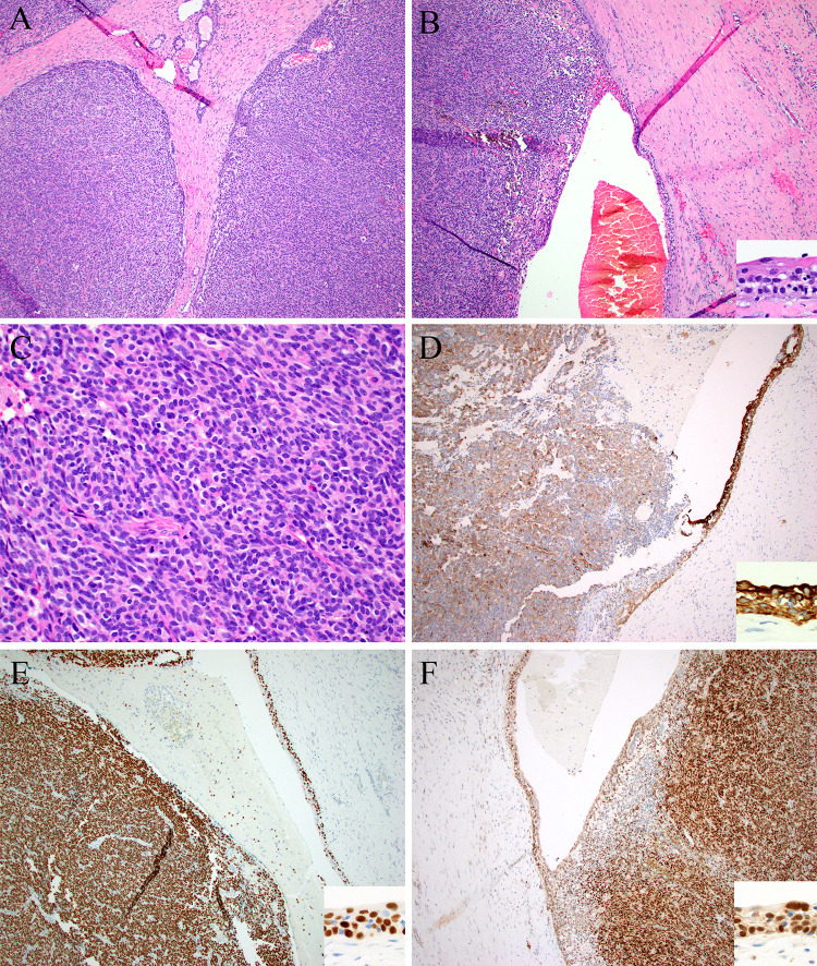
Fig. 3.
Case 3 was a large mass made up of solid nodules with foci of entrapped minor salivary glands (top) (A) and foci of cystic change lined by squamous epithelium (inset) (B). The tumor cells were epithelioid to vaguely spindled, with uniform oval nuclei and pale eosinophilic cytoplasm (C). By immunohistochemistry, the solid and cystic elements (insets) were positive for pan-cytokeratin (D), p40 (E), and STAT6 (F)

