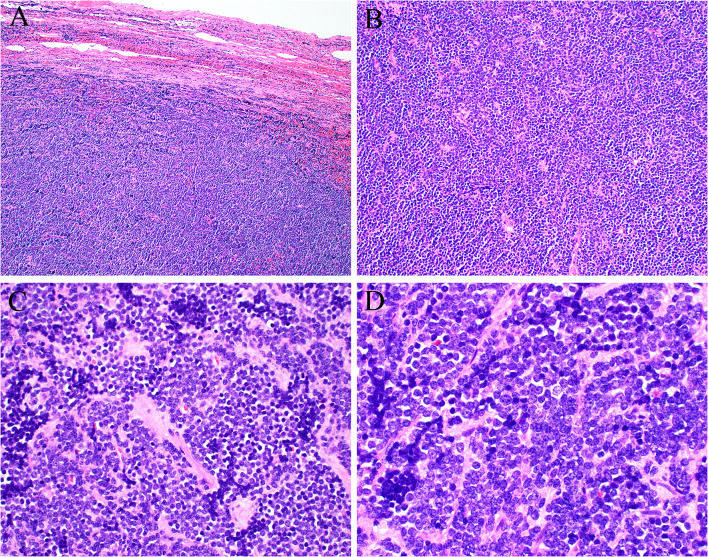
Fig. 2.
Excisional biopsy showed a lymph node with residual capsule and subcapsular sinus (A) otherwise obliterated by sheets of primitive round cells showing no signs of specific differentiation (B). The tumor cells had scant amounts of pale cytoplasm and uniform round nuclei, with small amounts of fibrotic stroma (C). The tumor nuclei were round to oval shaped with coarsely clumped chromatin (D)

