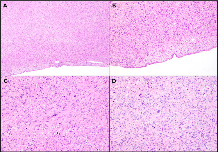
Fig. 2.
Histologic findings of leiomyosarcoma showing. A A subepithelial proliferation without involving the surface epithelium (H&E 4 ×). B Proliferation of spindle cells without involvement of the surface epithelium (H&E 10 ×). C Marked nuclear pleomorphism, hyperchromasia and numerous mitotic figures (H&E 20 ×). D Marked nuclear pleomorphism (H&E 20 ×)

