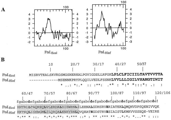
FIG. 3.
Comparison between E. coli and H. influenzae FtsL. (A) Kyte and Doolittle hydropathy plot comparison between FtsLEcol and FtsLHinf. (B) Amino acid sequence comparison between FtsLEcol and FtsLHinf. The first and second numbers indicated above the sequences correspond to amino acid positions in FtsLEcol and FtsLHinf, respectively. FtsLEcol and FtsLHinf membrane-spanning segments are in oversized boldface. The shaded area corresponds to the region exchanged in the LLHinfL swap (see text). The a to f positions of the heptad repeat of the potential coiled-coil domain are indicated above the sequence of the periplasmic domain of FtsLEcol and FtsLHinf. The d position is indicated in boldface. Conserved residues are indicated with a star below the sequence. Conservative substitutions are indicated with dots.

