Fig. 1.

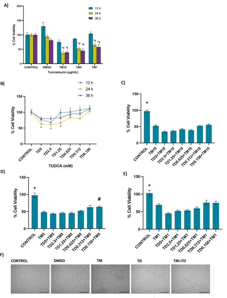
Cell viability assessed by MTT assay in HEK-293 cells. A) Dose-dependent 36-h time-course response analysis of tunicamycin-induced cell death. DMSO, cells treated with dimethyl sulfoxide (1 μl/ml); TM10, TM5, and TM1, cells treated with 10, 5, and 1 μg/ml tunicamycin. Data shown are representative of 7–8 separate experiments, and values are shown as mean ± SEM. Statistical analysis was performed by two-way ANOVA with all pairwise multiple comparison procedures done by Tukey multiple comparison test. *P < 0.05 vs. control and DMSO within the same incubation time. #P < .05, vs. 12 h within the same dose. No toxicity was observed at 12-h incubation in any TM doses. B) Dose-dependent effect of TUDCA (TD) on cell viability at 12–36-h time course. Data shown are representative of 7–8 separate experiments, and values are shown as mean ± SEM. Statistical analysis was performed by two-way ANOVA with all pairwise multiple comparison procedures done by Tukey multiple comparison test. *P < 0.05, vs. control within the 24-h incubation time. No toxicity was observed in cell viability at 12- and 36-h time-course compared to control. C) No protective effect was observed at 12-h TUDCA (TD) administration in 24 h 10 μg/ml TM toxicity. Control cells were incubated with only medium. HEK-293 cells were incubated with 10 μg/ml TM for 24 h. In the TM + TUDCA groups, TUDCA (.156–5 mM) was given 12 h after 10 μg/ml TM application, with a total TM incubation time of 24 h. Data shown are representative of 11–12 separate experiments, and values are shown as mean ± SEM. Statistical analysis was performed by one-way ANOVA with all pairwise multiple comparison procedures done by Dunnett’s T3 multiple comparison test. *P < 0.05 vs. all other groups. D) Protective effect of TUDCA (TD) was observed at 12 h .156 mM TD administration in 24 h 5 μg/ml TM toxicity. Control cells were incubated with only medium. HEK-293 cells were incubated with 5 μg/ml TM for 24 h. In the TM + TUDCA groups, TUDCA (.156–5 mM) was given 12 h after 5 μg/ml TM application, with a total TM incubation time of 24 h. Data shown are representative of 11–12 separate experiments, and values are shown as mean ± SEM. Statistical analysis was performed by one-way ANOVA with all pairwise multiple comparison procedures done by Dunnett’s T3 multiple comparison test. *P < 0.05, vs. all other groups. #P < 0.05, vs. TM5. E) No protective effect of TUDCA (TD) was observed at 12-h TD administration in 24 h 1 μg/ml TM toxicity. Control cells were incubated with only medium. HEK-293 cells were incubated with 1 μg/ml TM for 24 h. In the TM + TUDCA groups, TUDCA (.156–5 mM) was given 12 h after 1 μg/ml TM application, with a total TM incubation time of 24 h. Data shown are representative of 11–12 separate experiments, and values are shown as mean ± SEM. Statistical analysis was performed by one-way ANOVA with all pairwise multiple comparison procedures done by Dunnett’s T3 multiple comparison test. *P < 0.05, vs. all other groups. F) Morphological changes of TM and TUDCA administration on HEK-293 cells observed under an inverted light microscope (20X magnification) bar, 100 μm. Cells treated with 1 μl/ml DMSO for 24 h or .156 mM TUDCA for 12 h showed no significant morphological changes. Cells treated with 24 h, 5 μg/ml TM showed changes such as shrinkage, aggregation, detachment, and rounding. A decrease in cell population was also noted in the TM group. Administration of .156 mM TUDCA after 12 h 5 μg/ml TM showed a decrease in TM-related morphological deterioration.
