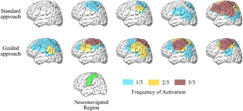
Fig. 3.
fNIRS reproducibility with a neuronavigation system. Frequency of activated brain regions during a motor task for the standard and guided approaches for probe positioning across five participants. The standard procedure used a tape to record head-size and find the optode location relative to the 10-20 system, while the guided approach used real-time neuronavigation software to place the optodes on the target region of interest (motor cortex, shown in green in the reference brain located at the bottom of the figure). Data were collected at three sessions on three different days for every subject. The frequency of activation represents reproducibility.

