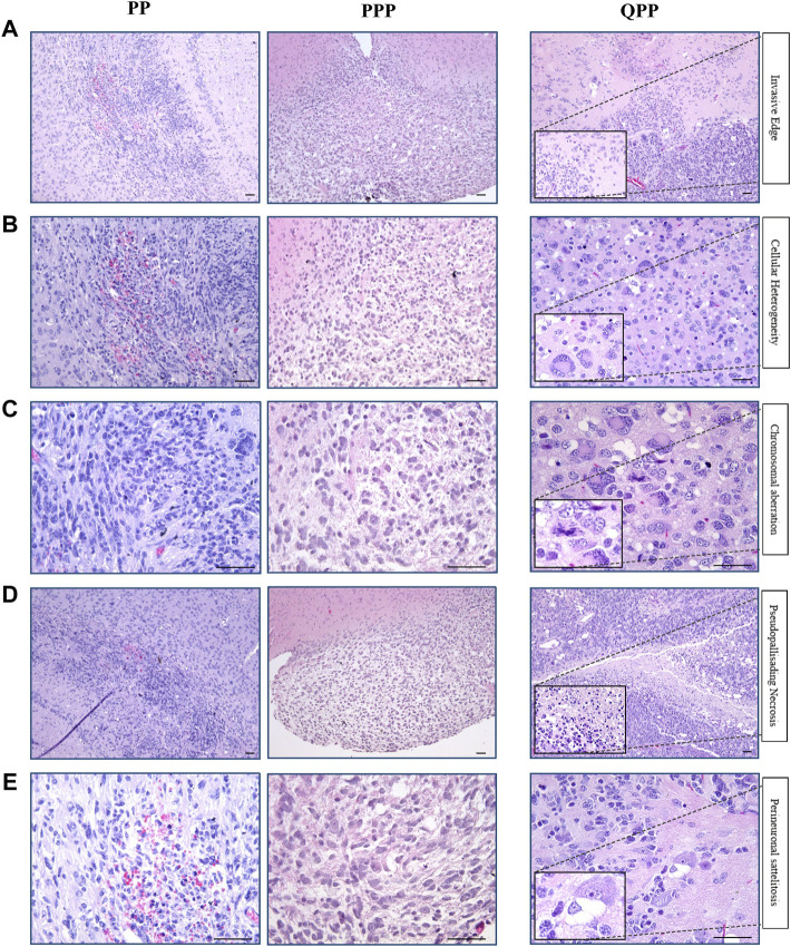
FIGURE 3.
Histopathological analyses identified the brain tumor isolated from the PPP cohort as low-grade glioma. (A). Representative H&E images of tumors harvested from QPP, PPP, and PP cohorts demonstrating invasive edges. (B). Representative H&E images of tumors harvested from QPP, PPP, and PP cohorts indicating intra-tumor cellular heterogeneity. (C). H&E images of tumors harvested from QPP, PPP, and PP cohorts representative of chromosomal aberrations. (D). Representative H&E images of tumors harvested from QPP, PPP, and PP cohorts displaying intra-tumor necrosis. (E). Representative H&E images of tumors harvested from QPP, PPP, and PP cohorts exemplifying perineuronal satellitosis. Scale bars represent 50 μm.

