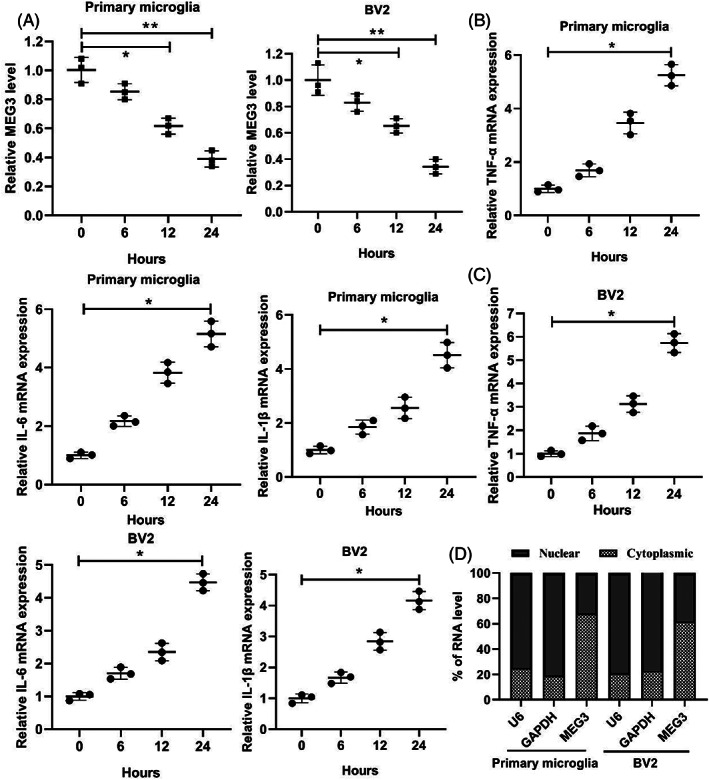
FIGURE 2.

The changes of lncRNA MEG3 expression in the spinal cord injury model in vitro. (A) Primary mouse microglia were isolated from the normal mouse and cultured, and mouse microglia cell line BV2 was also cultured, and then treated the cells with 1 μg/ml LPS for 0, 6, 12, and 24 h. qRT‐PCR was applied to test the expression of lncRNA MEG3. (B) qRT‐PCR was applied to detect the expressions of TNF‐α, IL‐6, and IL‐1β in the primary microglia. (C) qRT‐PCR was applied to detect the expressions of TNF‐α, IL‐6, and IL‐1β in the BV2 cells. (D) The distribution of lncRNA MEG3 in mouse primary microglia and BV2 cells by qRT‐PCR. *P < 0.05, **P < 0.01 vs. Control (0 h). Data are represented as the mean ± SD of three independent assays
