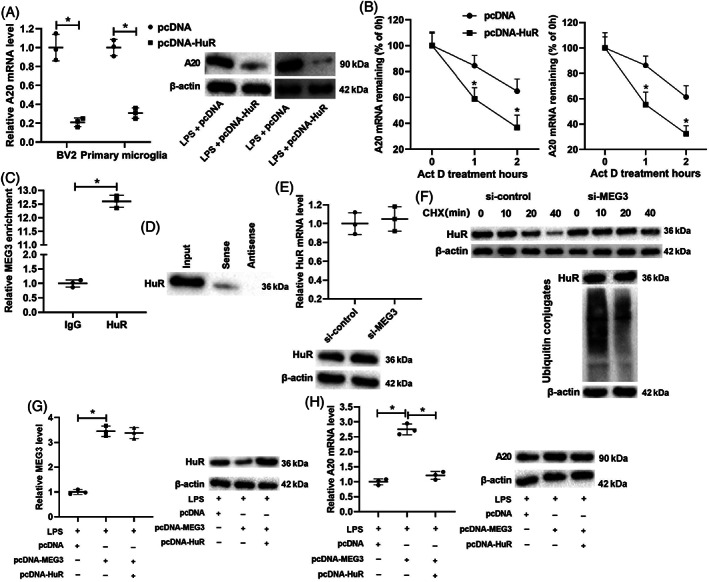
FIGURE 5.

HuR regulates the degradation of A20 mRNA and verification of the interaction between lncRNA MEG3 and HuR protein. (A) pcDNA‐HuR was transfected into BV2 cells and primary microglia for 48 h and then treated the cells with LPS for 24 h. qRT‐PCR and western blot were applied to test the mRNA and protein levels of A20. (B) pcDNA‐HuR was transfected into BV2 cells and primary microglia and then treated cells with an RNA synthesis inhibitor Actinomycin D for 0, 1, and 2 h. qRT‐PCR was performed to measure the mRNA level of A20. (C,D) RNA immunoprecipitation (RIP) and RNA pull‐down assays in BV2 cells were conducted to confirm the interaction between lncRNA MEG3 and HuR protein. (E) si‐MEG3 was transfected into BV2 cells. qRT‐PCR and western blot were applied to test the mRNA and protein levels of HuR. (F) si‐MEG3 was transfected into BV2 cells and then treated cells with a protein synthesis inhibitor cycloheximide (CHX) for 0, 10, 20, and 40 min. CHX‐chase and ubiquitination assays were applied to analyze the ubiquitination degradation of the protein. (G) pcDNA‐MEG3 and/or pcDNA‐HuR was transfected into BV2 cells, and the cells were treated with LPS for 24 h. qRT‐PCR was applied to measure the expression of lncRNA MEG3, and western blot was performed to test the protein level of HuR. (H) qRT‐PCR and western blot were conducted to test the mRNA and protein levels of A20. *P < 0.05 vs. LPS + pcDNA, pcDNA, IgG, or LPS + pcDNA‐MEG3. Data are represented as the mean ± SD of three independent assays
