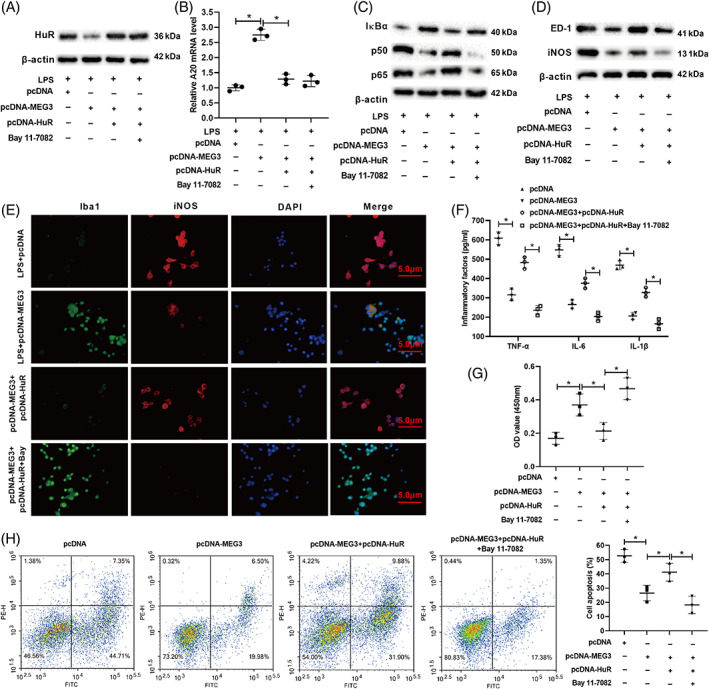
FIGURE 6.

lncRNA MEG3 affects the M1 polarization of microglia through the HuR/A20/NF‐κB axis. (A) pcDNA‐MEG3 and/or pcDNA‐HUR was transfected into BV2 cells and the cells were treated with an NF‐κB pathway inhibitor BAY 11‐7082, and the cells were further treated with LPS for 24 h. Western bolt was applied to detect the protein level of HuR. (B) qRT‐PCR was applied to measure the mRNA level of A20. (C,D) Western bolt was applied to test the protein levels of IκBα, p50, p65, ED‐1, and iNOS. (E) Immunofluorescence assay was applied to analyze the expressions of Iba‐1 and iNOS (scale bar: 5 μm). (F) ELISA was applied to detect the concentrations of TNF‐α, IL‐6, and IL‐1β. (G) BV‐2 cells were co‐incubated with human nerve cells SH‐SY5Y for 12 h. CCK‐8 was applied to assess the proliferation of SH‐SY5Y cells. (H) Flow cytometry assay was applied to analyze the apoptosis of SH‐SY5Y cells. *P < 0.05 vs. LPS + pcDNA, LPS + pcDNA‐MEG3, pcDNA, or pcDNA‐MEG3 + pcDNA‐HuR. Data are represented as the mean ± SD of three independent assays
