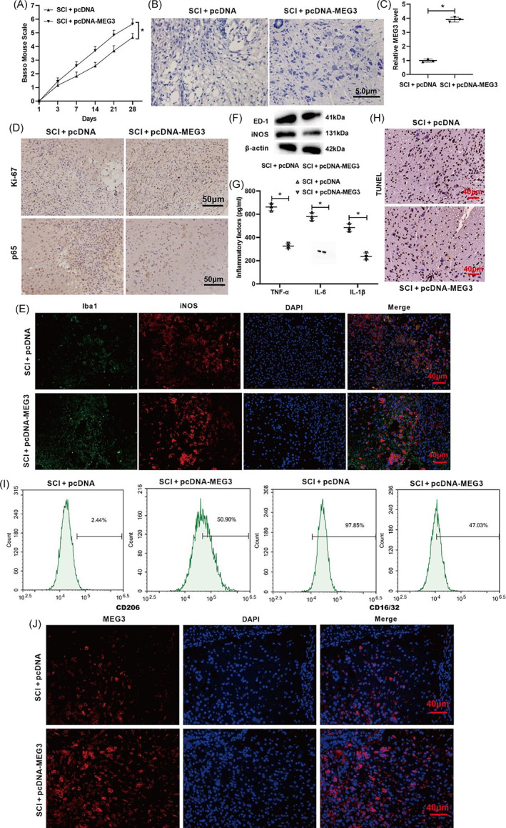
FIGURE 7.

Influence of lncRNA MEG3 on motor function recovery and neuroinflammation relief in SCI mice. (A) The mouse model of SCI was constructed and pcDNA‐MEG3 was injected intrathecally into mice. BMS score was performed to analyze the motor function of mice. (B) Nissl staining was conducted to assess the pathological characteristics of spinal cord tissues in mice (scale bar: 5 μm). (C) qRT‐PCR was conducted to test the expression of lncRNA MEG3. (D) The immunohistochemical assay was applied to detect the expressions of Ki‐67 (a marker for proliferation) and p65 (scale bar: 50 μm). (E) Immunofluorescence was applied to analyze the expressions of Iba1 and iNOS (scale bar: 40 μm). (F) Western blot was performed to test the protein levels of ED‐1 and iNOS. (G) ELISA was applied to measure the concentrations of TNF‐α, IL‐6, and IL‐1β. (H) TUNEL assay was conducted to assess the cell apoptosis (scale bar: 40 μm). *P < 0.05 vs. SCI + pcDNA. SCI, spinal cord injury. (I) Primary mouse microglia were isolated from the normal mouse and cultured. CD206 (M2) and CD16/32 protein expressions were measured by flow cytometry assay. (J) RNA fluorescence in situ hybridization (RNA FISH) assay was used to detect MEG3 expression in mouse spinal cord tissues. Data are represented as the mean ± SD of three independent assays [Correction added on 1 August 2022, after first online publication: Figure 7 has been replaced.]
