
FIGURE 2.
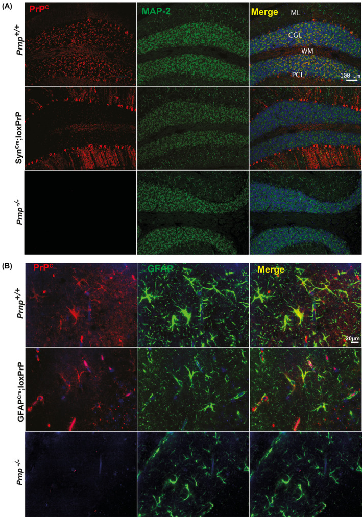
PrPC expression patterns in CAG‐CAT‐PrP mice. (A and B) Cerebellar sections of wild‐type and transgenic mice (as indicated) were immunostained for PrPC (red) as well as for MAP2 (microtubule‐associated protein 2), GFAP (glial fibrillary acidic protein), which were used as neuronal and astrocytic markers, respectively (green). As controls cerebellar sections from Prnp ablated mice were used. Blue: cell nuclei (DAPI). In Prnp +/+ mice, PrPC is detected in the cerebellar granule cell layer (CGL), Purkinje cells (PCL), and molecular layer (ML). In SynCre;loxPrP cerebella (A), PrPC was mostly detected in Purkinje cells. In GFAPCre;loxPrP, PrPC was exclusively detected in astrocytes colocalizing with GFAP. Prnp ablated mice revealed absence of any unspecific staining. Each staining was performed on sections from at least three individual mice