
FIGURE 3.
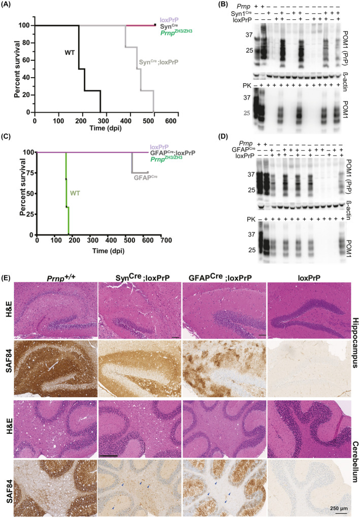
Neuron‐selective, but not astrocyte‐selective, PrPC expression confers susceptibility to prion disease. (A) Survival of SynCre;loxPrP, loxPrP, SynCre, Prnp ZH3/ZH3 and wild‐type mice inoculated intracerebrally with RML6 prions. The median incubations for SynCre−;loxPrP (n = 5) and wild‐type mice (n = 4) were 435 and 195 days post inoculation (dpi), respectively. Survival curves were compared by a log‐rank (Mantel‐Cox) test. (B) Total PrP (upper panel) and PK‐resistant PrPSc (lower panel) in brains of prion‐infected, terminally sick mice. SynCre;loxPrP and wild‐type brains, but not loxPrP brains, contained PrPSc. β‐actin: loading control. Lane #2 (upper panel) was intentionally underloaded to avoid overexposure. (C) Survival curves of GFAP1Cre;loxPrP, loxPrP, Prnp ZH3/ZH3 and wild‐type mice inoculated with RML6 prions intracerebrally. GFAPCre;loxPrP (n = 5) did not develop clinical signs of prion disease. Wild‐type mice (n = 4): 167 dpi (median incubation time). One of the GFAPCre mice was sacrificed (544 dpi) because of acute dermatitis and did not exhibit scrapie signs or PrPSc accumulation. (D) Western blot of total PrP and PK‐digested PrPSc in brain homogenates of prion‐infected GFAPCre;loxPrP (627 dpi) and control mice. GFAPCre;loxPrP harbor copious PrPSc. (E) Hippocampal and cerebellar histology of prion‐infected SynCre;loxPrP, GFAPCre;loxPrP, loxPrP, and Prnp +/+ mice. Slices were stained with hematoxylin and eosin (H&E) and anti‐PrP antibody SAF84. Both astrocyte‐ and neuron‐restricted PrP transgenic mice accumulated PrPSc, but their deposition patterns differed profoundly. Blue arrows in cerebellar regions of SynCre;loxPrP and GFAPcre;loxPrP: PrPSc deposits