FIGURE 1.

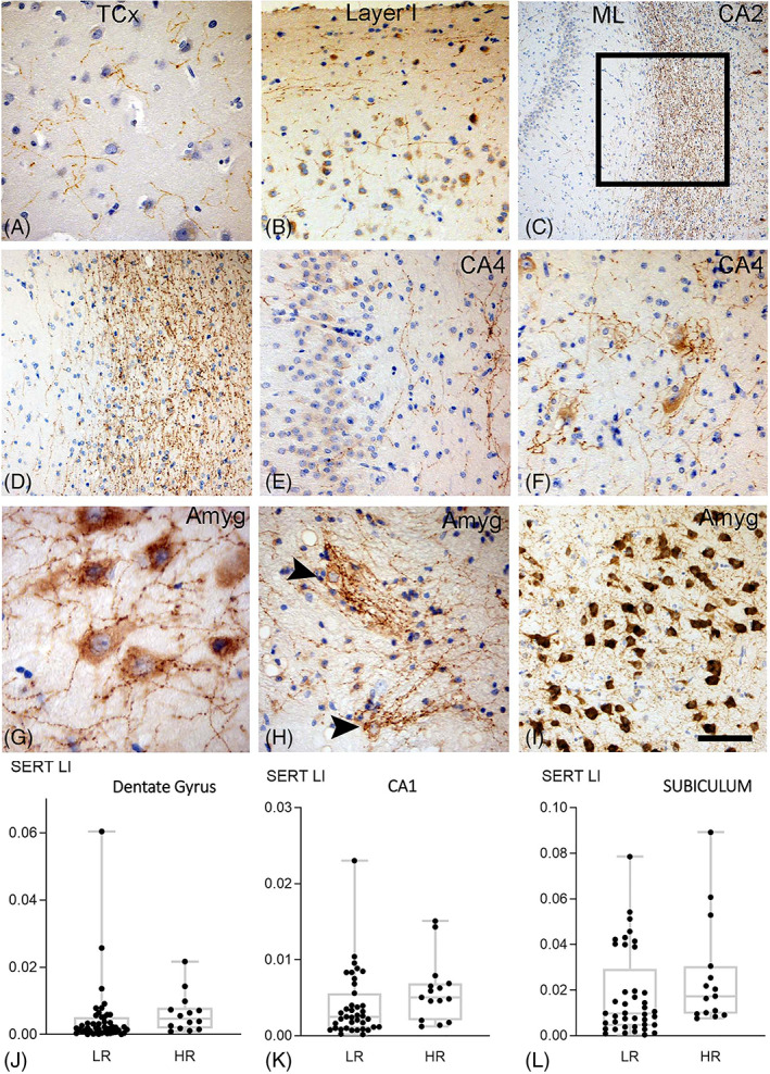
SERT in TLE surgical series with hippocampal sclerosis. (A) Fine axonal varicose SERT networks were present in temporal neorcortex (TCx). (B) There was an impression of condensation of SERT‐positive fibres in the superficial cortical layers (Layer I). (C) Dense SERT axonal networks were also present in the hippocampal white matter (stratum radiatum, lacunosum and moleculare), shown here between the molecular layer (ML) of the dentate gyrus and the pyramidal cell layer of CA2 (box shown at higher magnification in (D)) (E) SERT axons were also present in the dentate gyrus and CA4 region. (F) In CA4 ‘nets’ of SERT positive processess surrounded pyramidal neurones. (G) The amygadala (Amyg) was enriched in SERT with numerous beaded axons in proximity to neurones. (H) SERT positive neurones with complexed tuft like branches were also present in the amygdala (Amyg). (I) Amygdala regions with intense SERT positivity were observed. Scatter plots of mean SERT labelling index (LI) in high risk (HR) compared to and low risk (LR) for SUDEP cases which showed a significant increase in the high risk group in the (J) dentate gyrus, (K) CA1 and (L) subiculum regions. Bar in I equivalent to approx. 50 microns in A, B. D. E. F, H, I, 150 microns in C and 35 microns in H
