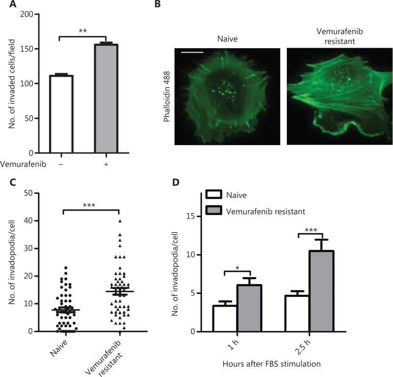
Figure 1.
Vermurafenib resistant melanoma cells exhibited more invasiveness and invadopodia formation. (A) Quantitation of the invaded cells in vemurafenib resistant WM793 cells cultured with and without vemurafenib (n = 3). (B, C) Representative images of actin fluorescence staining showing invadopodia (B) and quantification of invadopodia (C) in naïve and vermurafenib resistant WM793 cells (n = 3). (D) Quantitation of invadopodia at indicated times after 10% fetal bovine serum stimulation in naive and vermurafenib resistant WM793 cells (n = 3). Scale bars = 10 μm. *P < 0.05; **P < 0.01; ***P < 0.001.

