Figure 3.
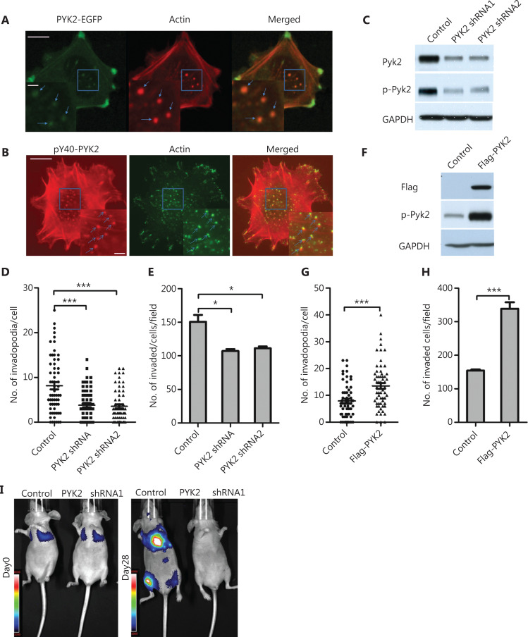
Phospho-PYK2-Y402 localizes at invadopodia and regulates invadopodia formation and cell invasion in naïve WM793 cells. (A) Representative images of fluorescence staining shows that PYK2-EGFP was localized at invadopodia (actin staining). (B) Representative images of fluorescence staining show p-PYK2 localized at invadopoida. (C–E) Western blot analysis of expressions of PYK2 and p-PYK2 (C), quantification of invadopodia (D) and invaded cells (E) in PYK2-knockdown and control WM793 cells (n = 3). (F–H) Western blot analyses of PYK2 and p-PYK2 expressions in PYK2-transfected and control WM793 cells (F), and quantification of invadopodia (G) and invaded cells (H) in PYK2-overexpressing and control WM793 cell (n = 3). (I) Knockdown of PYK2 in 1205Lu cells significantly reduced lung metastases in a mouse model. Scale bars = 10 μm in the main images; Scale bars = 2 μm in the inserts. *P < 0.05; ***P < 0.001.

