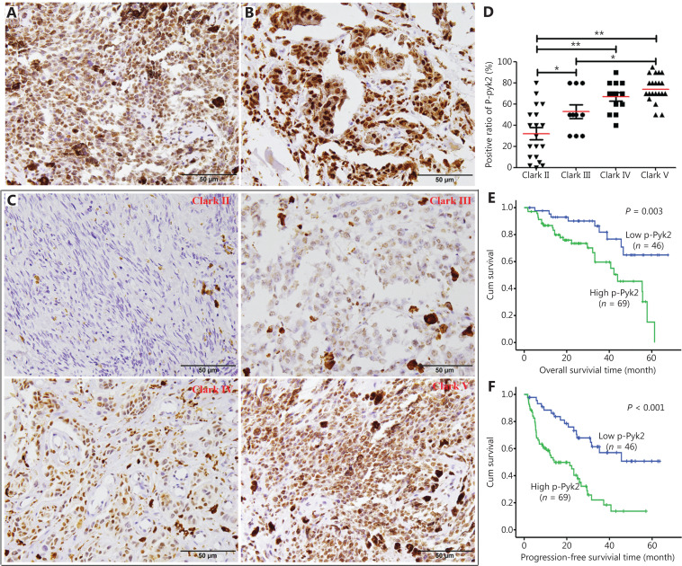
Figure 7.
Expression of PYK2-Y402 in melanoma tissues and survival analysis of melanoma patients. (A, B) Representative images of p-PYK2 immunohistochemical staining in lymph node metastasis (A) and distant metastasis (B) of melanomas. (C) Representative images of p-PYK2 immunohistochemical staining of Clark grade II, III, IV, and V melanomas. (D) Comparison analyses of p-PYK2 expression levels in melanomas with different Clark grades. (E) The curves of overall survival according to p-PYK2 expressions in patients with melanomas. (F) The curves of progression-free survival according to p-PYK2 expressions in patients with melanomas. *P < 0.05; **P < 0.01.

