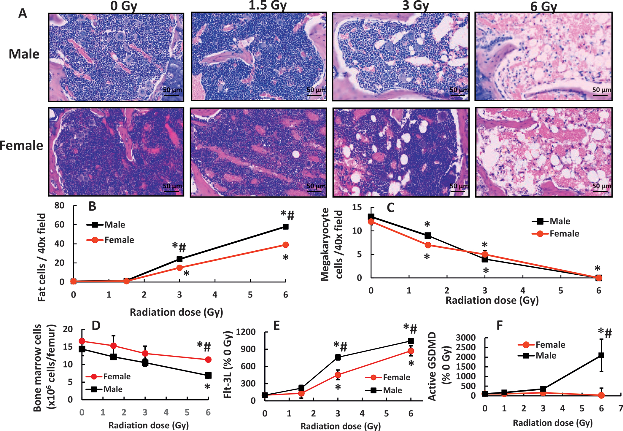
FIG. 1.

Mixed-field (67% neutron + 33% gamma) radiation injures bone marrow morphology more in male mice than in female mice. Bone marrow histology slides on day 7 post-mixed-field irradiation stained with hematoxylin and eosin (panel A). N = 4 per group. Fat cells (panel B) and megakaryocyte counts (panel C). N = 4 per group, 4 fields/slide with ×40 magnification were measured. Panel D: Bone marrow cells collected from femurs were counted. N = 4 per group. Panel E: Flt-3 ligand concentrations in blood were measured (N = 4 per group). Panel F: Active gasdermin D (GSDMD) was measured in bone marrow cell lysate from femurs (N = 3–4 per group). Data are shown as mean ± sem except Flt-3 ligand data which were mean ± sd. *P < 0.05 vs. 0 Gy; #P < 0.05 male vs. female.
