Figure 1.
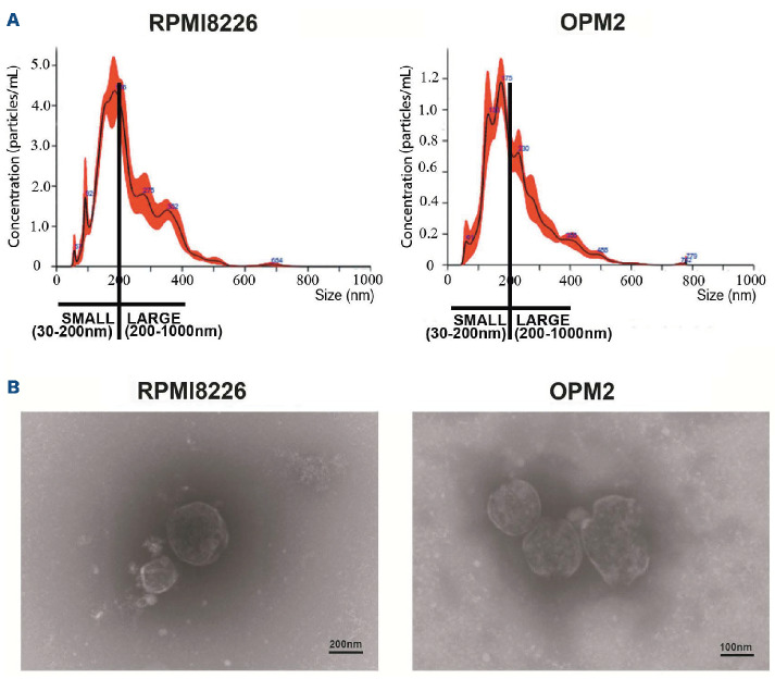
Characterization of multiple myeloma cell-released extracellular vesicles. Extracellular vesicles (EV) from multiple myeloma cell lines (HMCL) RPMI8226 and OPM2 cells (MM-EV), were isolated by ultracentrifugation and analyzed by (A) nanot-racking particle analysis (NTA) and (B) electron transmission microscopy (TEM). (A) NTA analysis reveals the presence of small (30-200 nm) and large (200-1,000 nm) vesicles. Size and concentration of EV were determined by NanoSight NS300 system (Malvern Panalytical Ltd, Malvern, UK). A camera level of 12 and 5 30-second recordings were used for the acquisition of each sample of 3 independent EV isolations and one representative image is shown. (B) TEM analysis confirms the isolation of intact small and large vesicles.

