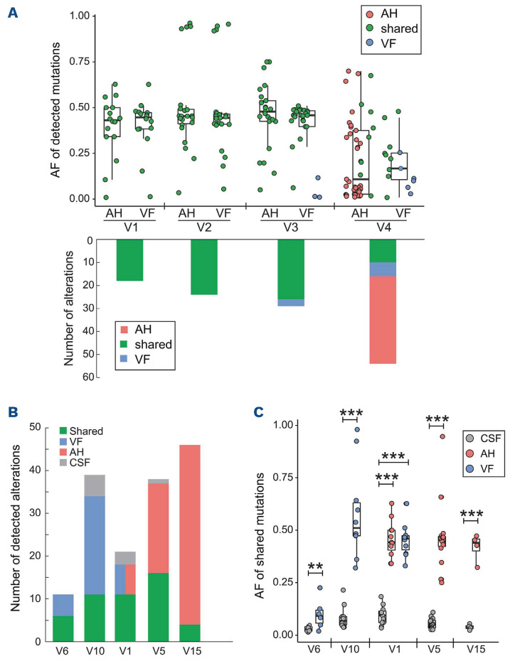
Figure 2.
Mutational comparisons between matched aqueous humor, vitreous fluid and cerebrospinal fluid samples. (A) Four pairs of baseline aqueous humor (AH) and vitreous fluid (VF) samples underwent targeted next-generation sequencing. The allele frequencies of shared and unique mutations detected in matched AH and VF samples are shown in the top panel. The number of alterations detected in each patient is shown at the bottom of the panel. Shared, AH-only, and VF-only alterations are colored green, red, and blue, respectively. (B) The number of alterations detected in cerebrospinal fluid (CSF) and AH/VF samples from five patients is as labeled in the legend. The brain involvement in patients V1 and V10 was confirmed by positron emission tomography/computed tomography examinations. (C) Allele frequencies of mutations present in both CSF and AH/VF samples. **P<0.05, ***P<0.01. AF: allele frequency.

