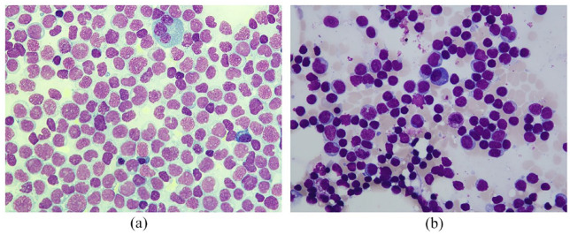
Figure 1.
Cytomorphology of bone marrow aspirates. (a) Bone marrow aspirate smear at time of diagnosis (50× magnification) showed subtotal infiltration of myeloid blasts replacing normal hematopoiesis without evidence of myeloid maturation, leading to diagnosis of acute myeloblastic leukemia without maturation according to WHO criteria. (b) Bone marrow aspirate smear at time of relapse (50× magnification) revealed persistence of cytomorphologic remission with a blast count below 5%, myeloid maturation, and normal findings for erythroid and megakaryocytic components. Bone marrow smears (a and b) were stained with the May–Grunwald–Giemsa kit and examined with the Nikon Eclipse E600 microscope. High-resolution pictures were taken with the mounted Nikon DSFi2 camera and processed with the Nikon Imaging Software Elements.

