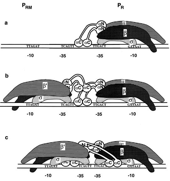
FIG. 4.
RNAP at pR interferes with open complex formation at pRM for the wild-type (a and b) but not the D10 (c) interpromoter distance. The α subunits of RNAP are shown in white, with the N-terminal domains anchored to the β and β′ subunits of the RNAP (gray and striped regions) and the CTDs and the flexible linkers jutting away from RNAP. The pRM promoter is on the left, and pR is on the right. The sequences of the −10 and −35 regions are indicated for the nontemplate strand of each promoter. The spacer DNAs between the −10 and the −35 regions are shown as devoid of contacts with RNAP. (a) The −35 regions of pR and pRM are separated by 13 pdb. Within seconds of the addition of RNAP, an open complex forms at the pR promoter. Proposed upstream contacts of the α-CTDs of the RNAP are shown. (b) Subsequent interaction of RNAP with pRM in the presence of an RNAP at pR. The RNAP at pR obstructs upstream access by the α-CTDs of the RNAP at pRM. (c) The −35 regions of pR and pRM are separated by 2 bp. This closer-in arrangement allows the spacer DNAs of pR and pRM to be contacted by the α-CTDs of the RNAP at the other promoter, facilitating open complex formation at pRM despite the presence of an RNAP at pR.

