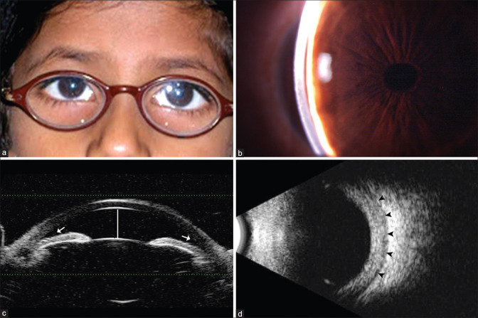
Figure 1.
(a) Clinical photograph of nanophthalmic child showing thick hyperopic glass, (b) Slit-lamp photo of the same child showing shallow anterior chamber depth, (c) UBM photo showing shallow anterior chamber (white line) and crowded angle structures (white arrow), (d) B-scan image showing increased retinochoroidal scleral thickness (black arrowheads)

