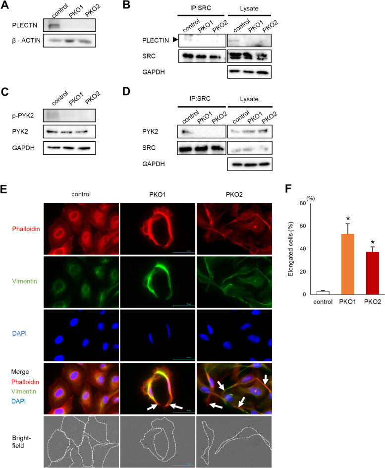
Fig. 1.
Src signaling is impaired in plectin deficient cells. A PLECTIN levels in control B16 and 2 clones of plectin deficient B16 cells (PKO1 and PKO2) were determined by western blotting analysis. B SRC was immunoprecipitated with an anti-Src antibody (IP:SRC) and interaction with PLECTIN was detected by western blotting analysis. C Expression and phosphorylation of PYK2, or GAPDH was determined by western blotting assay 24 h after plating. D Src was immunoprecipitated with an anti-Src antibody (IP:SRC) and interaction with PYK2 was detected by western blotting analysis. E Cells were stained with Rhodamine-phalloidin and vimentin/anti-mouse Alexa fluor 488. Long actin fibers are indicated by arrows. Cell shapes are outlined with a dotted line in the bright-field image. Scale bar = 50 μm. F Quantification of the number of elongated cells in samples shown in (E) (n = 5). *; p < 0.05 vs control

