Fig. 3.
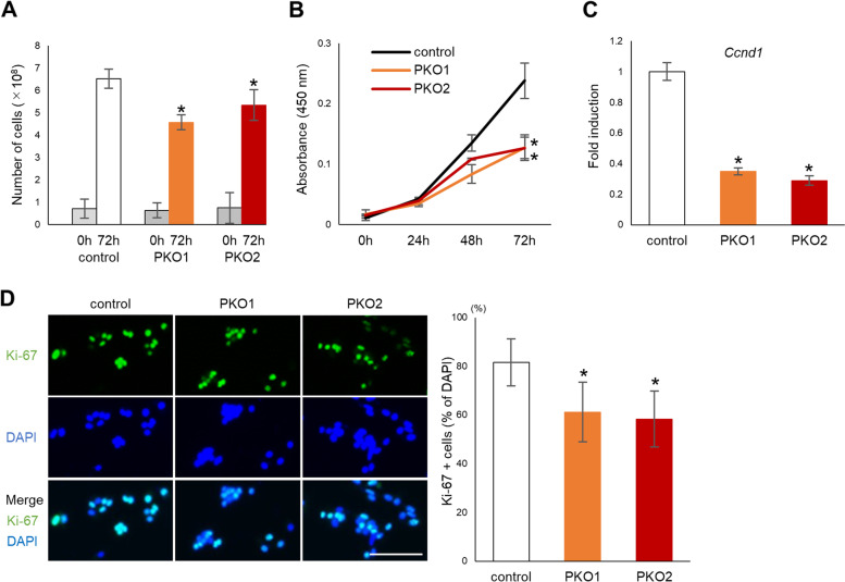
Cell proliferation is decreased in plectin knockout cells. A Cells were plated at a density of 2.5 × 10⁴ cells per well in 24-well plates for the indicated time period after which cells were trypsinized and the number of cells determined with an automated cell counter (n = 6). B Cells were plated at a density of 5 × 10³ cells per well in a 96 well plate and the number of viable cells measured by CCK-8 assay at the indicated time (n = 4). C mRNA was harvested from control or PKO cells 1 d after plating. Expression of Ccnd1 was determined by real time qPCR (n = 3). D Control or plectin knockout B16 cells were fixed and stained with anti-Ki-67/Alexa fluor 555 and DAPI. The percentage of Ki-67 positive cells relative to DAPI positive cells was calculated. (n = 5), *; p < 0.05 vs control

