Fig. 1.
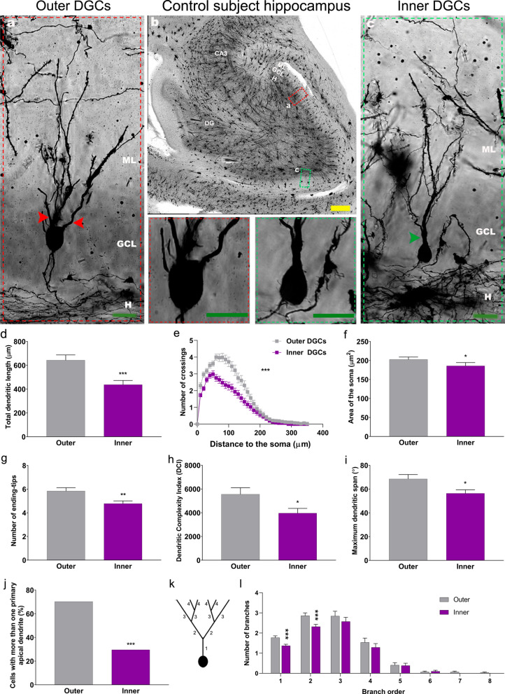
Morphological features of human dentate granule cells (DGCs) located in the outer and inner granule cell layer (GCL) of neurologically healthy control subjects. a-c Representative images of Golgi-stained human hippocampi and high-power magnification images showing the somata and primary dendrites of DGCs. d Total dendritic length. e Sholl´s analysis. f Area of the soma. g Number of ending-tips. h Dendritic complexity index (DCI). i Maximum dendritic span. j Percentage of cells with more than one apical primary dendrite. k Schematic representation of dendrite branch orders. l. Number of dendrites in each branch order. Yellow bar: 500 µm. Green bar: 20 µm. DG, dentate gyrus; GCL, granule cell layer; H, hilus; ML, molecular layer. Red and green arrowhead: apical primary dendrites. n = 127 cells obtained from 5 neurologically healthy control subjects. * 0.05 > P ≥ 0.01; ** 0.01 > P ≥ 0.001; and *** P < 0.001. Asterisks represent statistically significant differences in unpaired two-tailed Mann–Whitney U or Chi-squared test

