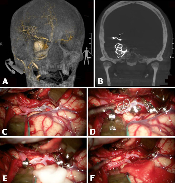
FIG. 2.

Three-dimensional reconstruction CT revealed a giant saccular intracranial aneurysm at the right cavernous part of the ICA, 3.1 × 3.2 × 2.9 cm (A) along with a migrated coil occlusion at the distal M1 segment of the right MCA (curved arrow, B). After Sylvian fissure splitting, intraoperative footage revealed an intravascular migrated coil visibly inside the right M1 segment of the MCA (C). After securing this MCA segment properly with temporary aneurysm clips at both distal and proximal ends, the coiling material was retrieved through a small linear incision on the MCA (D and E). This MCA segment was later repaired by an interrupting simple suture using nylon suture material under a microscope (F).
