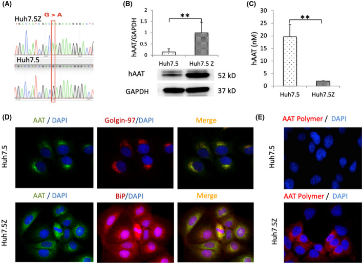
FIGURE 1.

AAT accumulation in Huh7.5Z cells. (A) Direct DNA sequencing showed a point mutation (G > A) in the human SERPINA1 gene at position 342 in Huh7.5Z cells. (B) Western blot analysis of AAT in cell lysate showed a significantly high level of intracellular AAT in Huh7.5Z cells compared with Huh7.5 cells (6.7‐fold). Results were normalized to GAPDH levels. Western blot quantification data are presented as mean ± SD. Significance was determined by the Student t test. **p < 0.01. (C) Enzyme‐linked immunosorbent assay results showed low AAT concentration in the cell culture medium of Huh7.5Z cells compared with that of Huh7.5 cells (0.1‐fold). Data are presented as mean ± SD. Significance was determined by the Student t test. **p < 0.01. (D) Immunofluorescence images (magnification, 40×) showed the colocalization of AAT (green) with golgin‐97 (red), a Golgi marker, in Huh7 and the colocalization of AAT (green) with BiP (red), an ER marker, in Huh7.5Z cells. AAT was mainly distributed in a Golgi pattern in Huh7.5 cells but an ER pattern in Huh7.5Z cells. (E) Immunofluorescence images (magnification, 40×) showed AAT polymer (red) in Huh7.5Z cells but not in Huh7.5 cells. AAT, alpha‐1 antitrypsin; BiP, binding protein; DAPI, 4′,6‐diamidino‐2‐phenylindole; ER, endoplasmic reticulum; GAPDH, glyceraldehyde 3‐phosphate dehydrogenase; hAAT, human alpha‐1 antitrypsin; SERPINA1, serpin family A member 1
