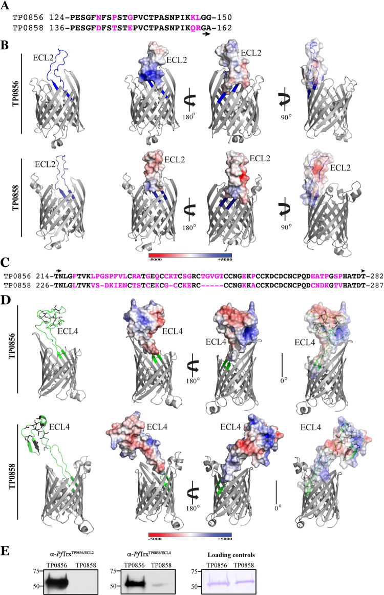
FIG 4.
Comparison of sequences, structures and electrostatics of ECL2 and ECL4 of TP0856 and TP858 (Nichols). (A) Alignment of TP0856 and TP0858 ECL2 sequences with substitutions shown in magenta. (B) trRosetta models of TP0856 and TP0858 with ECL2 shown in blue (left). Electrostatics of ECL2s are shown in the same and opposite orientations (middle and right, respectively). (C) Alignment of TP0856 and TP0858 ECL4 sequences with substitutions and deletions shown in magenta. (D) trRosetta models of TP0856 and TP0858 with ECL4 shown in blue (left). Electrostatics of ECL4s are shown in the same and opposite orientations (middle and right, respectively). Some ECLs and the hatches are masked for optimal viewing of electrostatics. (E) Immunoblot reactivity of rabbit anti-PfTrxTP0856/ECL2 and anti-PfTrxTP0856/ECL4 against full-length TP0856 and TP0858.

