FIG 1.
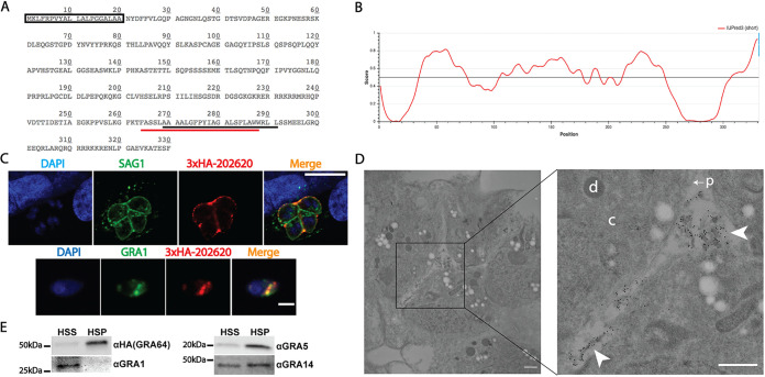
GRA64 is a membrane-bound dense granule protein localizing to the intravacuolar network during tachyzoite infection. (A) Amino acid sequence of TGME49_202620. The predicted signal peptide is indicated by a box (SignalP 5.0 prediction), while the predicted transmembrane domains are indicated by underlines (black, TMHMM prediction; red, Phobius prediction). (B) Results from IUPred3 short disorder prediction of TGME49_202620 protein intrinsic disorder are shown using medium smoothing (an approach within IUPred3 used to reduce the noise of amino acid residue free energy predictions based on the predictions of neighboring residues). Score indicates regions of low and high disorder. Low predicted disorder correlates with the signal peptide and transmembrane domain regions of the amino acid sequence indicated on the graph. (C) Top panel: IFA image of N terminus 3×HA epitope-tagged TGME49_202620 protein from a tachyzoite vacuole 1 day postinfection. TGME49_202620 is detected within the parasitophorous vacuole outside the parasite boundaries (ascertained by SAG1 labeling), with partial labeling of the parasitophorous vacuolar membrane (PVM). Scale bar = 10 μm. Bottom panel: immunofluorescence assay (IFA) images of an extracellular parasite expressing N terminus 3×HA epitope-tagged TGME49_202620 protein. TGME49_202620 (here, GRA64) colocalizes with GRA1. Scale bar = 5 μm. (D) Immunoelectron microscopy image of a tachyzoite vacuole expressing N terminus 3×HA epitope-tagged GRA64 protein, with select compartments labeled in the higher magnification inset image as follows: “d,” parasite dense granule; “c,” parasite cytoplasm; “p” with arrow, parasite plasma membrane; arrowheads, gold particle-labeled tubular material within the lumen of the parasitophorous vacuole. The 10-nm gold particles conjugated to anti-HA antibody appear to label the membranous structures of the intravacuolar network (IVN) within the parasitophorous vacuole lumen. The parasitophorous vacuole membrane was likely compromised by Triton X-100, which was used to permeabilize membranes for the labeling of GRA64 in this experiment. Bars, 500 nm. (E) Immunoblot images of GRA64, GRA1, GRA5, and GRA14 probed in high-speed supernatant (HSS, soluble) and high-speed pellet (HSP, membranous) fractions. GRA1, a soluble protein, is detectable only in the HSS fraction, whereas GRA64 is detectable predominately in the HSP fraction, similar to the previously described membrane-bound proteins GRA5 and GRA14.

