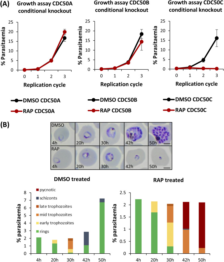
FIG 2.
(A) Growth curves showing parasitemia as measured by flow cytometry of CDC50A-HA:loxP, CDC50B-HA:loxP, and CDC50C-HA:loxP parasites treated with DMSO (vehicle-only control) or RAP. Means of results from three independent experiments are plotted. Error bars, standard deviations (SD). (B) Upper panel, Giemsa-stained thin blood films showing development of ring stage parasites following egress of synchronous DMSO- and RAP-treated CDC50C-HA:loxP schizonts. Ring formation occurs in RAP-treated CDC50C-HA parasites, but the parasites did not develop beyond the early trophozoite stage and eventually collapsed into small vacuoles. Scale bars, 2 μm. Lower panel, microscopic quantification of parasite developmental stages at each time point. RAP-treated parasites displayed an accumulation of pycnotic parasites at late life cycle stages and showed no expansion in parasitemia. Counts are means of results of two independent experiments.

