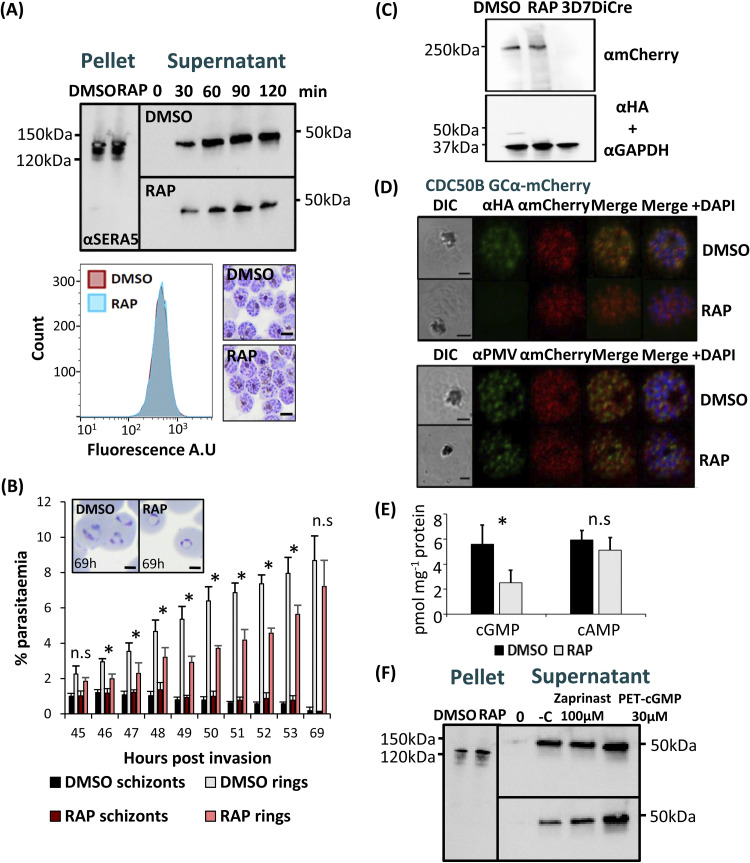
FIG 4.
(A) Western blot analysis monitoring egress kinetics of DMSO- and RAP-treated CDC50B-HA:loxP schizonts. Reduced detection of the SERA5 p50 proteolytic fragment in culture supernatants of RAP-treated CDC50B-HA:loxP parasites indicates an impaired egress rate in the absence of CDC50B-HA. Lower panel left, histograms of DNA (SYBR green) staining of DMSO- or RAP-treated schizonts. A total of 10,000 cells were counted per treatment. The image is representative of three independent experiments. Lower panel right, Giemsa-stained thin blood films of Percoll-purified DMSO- and RAP-treated schizonts. No delay in schizont maturation is evident in RAP-treated parasites. Images are representative of three independent experiments. Scale bar, 5 μm. (B) Flow cytometry analysis of ring formation by DMSO- and RAP-treated CDC50B-HA:loxP parasites. Samples from highly synchronized cultures treated at the ring stage were taken in triplicate at hourly intervals from 45 to 53 h and at 69 h postinvasion were stained with the DNA stain SYBR green. Samples were analyzed by flow cytometry, and the schizont and ring parasitemias were determined by gating high-signal and low-signal SYBR-positive cells, respectively. Mean parasitemia values (starting schizontemia adjusted to 2%) from two independent experiments are plotted. Error bars, SD; n.s, not significant; *, P < 0.05, by Student's t test for comparison of ring parasitemias between DMSO and RAP samples. Insets are smears taken from cultures at 69 h postinvasion. Scale bars, 2 μm. (C) Western blot analysis of DMSO- and RAP-treated CDC50B-HA:loxP GCα-mCherry and control 3D7DiCre schizonts. Top panel, ~250-kDa fragment detected by an mCherry antibody that is absent from control (untagged) schizont lysates. Lower panel, the same samples probed with an anti-HA antibody and an anti-GAPDH (PF3D7_1462800) loading control antibody. (D) Top, localization by IFA of CDC50B and GCα mCherry in RAP- and DMSO-treated CDC50B-HA:loxP GCα-mCherry schizonts. Bottom, localization of GCα mCherry and PMV (PF3D7_1323500), an ER marker in RAP- and DMSO-treated schizonts. Scale bar, 2 μm. (E) Quantification of cyclic nucleotide levels in tightly synchronized DMSO- and RAP-treated mature CDC50B-HA:loxP schizonts by direct ELISA. Means of results from three independent experiments are plotted. Error bars, SD; n.s, not significant; *, P < 0.05, Student's t test. (F) Restoration of egress of RAP-treated CDC50B-HA:loxP schizonts by treatment with zaprinast or PET-cGMP. Supernatant and pellet samples were taken at time point 0 after washing with RPMI 1640 medium, to control for parasite numbers and egress. Samples were then taken at 60 min postincubation at 37°C. Lane −C, no treatment. The image is representative of three independent experiments.

