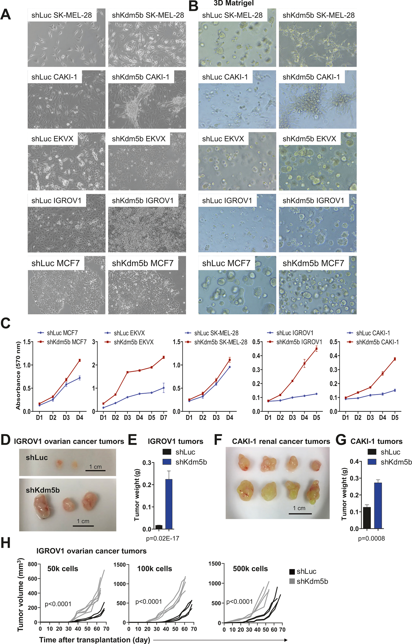
Fig. 6. Functional and morphological characteristics of KDM5B depleted cancer cells.

A Cancer cell lines transduced with shLuc or KDM5B-shRNA-2 (shKDM5B) lentiviral particles. shKDM5B CAKI-1, EKVX, and IGROV1 cancer cells exhibit morphological differences. KDM5B depleted CAKI-1 cells exhibit 3D growth characteristics, while KDM5B depleted EKVX are morphologically more homogenous, and “shiny” or circular IGROV1 cells became more prevalent upon knockdown of KDM5B. B shLuc and shKDM5B cancer cells cultured in 3D Matrigel. C MTT assays revealed that KDM5B-depleted cancer cells exhibit increased proliferation. Tumor growth assay. 106 shLuc or shKDM5B (D, E) IGROV1 or (F, G) CAKI-1 cells were subcutaneously injected into SCID-beige mice (n = 3 for IGROV1; n = 4 for CAKI1). E Weight of IGROV1 and G CAKI-1 tumors. Tumor weight plots are presented as mean ± SEM. H Limiting dilution tumor growth assay. 50k, 100k, or 500k shLuc or shKDM5B IGROV1 cells were subcutaneously injected into NSG mice (n = 4). Tumor volume was measured twice weekly. Statistical significance was determined using two-way ANOVA.
